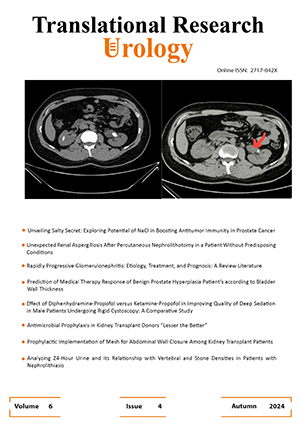
Computed Tomography in Pyonephrosis: The Diagnostic Performance of Hounsfield Unit Compared to Conventional Tools
نویسندگان
1 Isfahan Kidney Disease Research Center, Isfahan University of Medical Sciences, Isfahan, Iran
2 Isfahan Kidney Disease Research Center, Isfahan University of Medical Sciences, Isfahan, Iran
3 Department of Urology, School of Medicine, Isfahan University of Medical Sciences, Isfahan, Iran
4 Department of Urology, School of Medicine, Isfahan University of Medical Sciences, Isfahan, Iran
5 Department of Urology, School of Medicine, Isfahan University of Medical Sciences, Isfahan, Iran
6 Department of Urology, West Virginia University, Morgantown, WV, United States
doi
10.22034/tru.2025.512664.1234چکیده
Introduction: Pyonephrosis (PN) is a urological emergency that rapidly progresses to sepsis, requiring prompt diagnosis and drainage. Distinguishing PN from hydronephrosis (HN) remains a significant challenge in clinical practice. This study aimed to determine the optimal Hounsfield unit (HU) cut-off on non-contrast computed tomography (CT) scans and other CT features for accurate diagnosis of PN. Additionally, this study compared the diagnostic performance of CT with ultrasound.Methods: This prospective study included 80 patients undergoing nephrostomy placement. Clinical and laboratory data, ultrasound, and non-contrast CT scans were collected prior to the procedure. The turbidity of the aspirated fluid was recorded, and the specimen was sent for culture. The diagnostic accuracy of clinical, ultrasound, and CT parameters in predicting positive cultures and pus turbidity was analyzed.Results: Eighty patients were included in this study. The main ureteral obstructions were due to urolithiasis (53.8%), followed by bladder cancer (15.0%) and prostate cancer (11.3%). Among the patients, 42.5% had a positive culture, and 6.3% demonstrated highly turbid pus (class IV turbidity). An HU cut-off of 5 yielded 85.3% and 89.1% sensitivity and specificity in predicting the positive urine cultures, respectively (p<0.001). Furthermore, an HU threshold of 14.3 diagnosed pus turbidity class IV with 100% sensitivity and 89.3% specificity (p <0.001). Non-contrast CT also demonstrated superior diagnostic performance compared to ultrasound and other conventional clinical parameters.Conclusion: CT with HU estimation can reliably distinguish PN from HN, enhancing diagnosis and prompt treatment in such urological emergencies. Although CT outperformed conventional parameters in PUS detection through our study, further robust studies are required to draw strong evidence in this superiority.